Amygdala: Anatomy
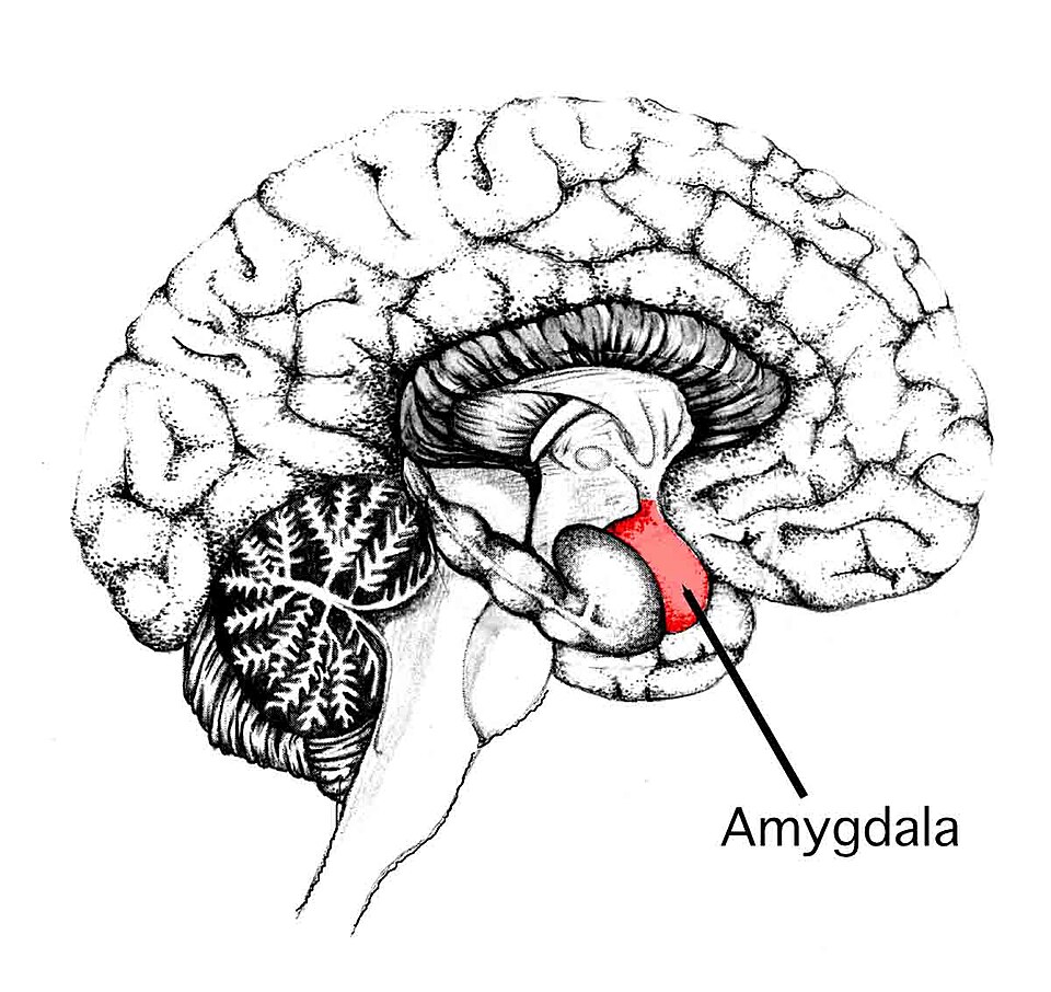
The amygdala is an almond-shaped collection of gray matter nuclei located in the anteromedial temporal lobe. It is a complex structure composed of multiple anatomically distinct nuclei rather than a single homogeneous mass.
The amygdala forms part of the limbic region of the telencephalon and occupies the anterior end of the temporal lobe, immediately anterior to the hippocampal formation.
Location
General Position
The amygdala is located:
- In the anterior temporal lobe
- Deep to the uncus
- Anterior to the hippocampus
- Inferomedial to the globus pallidus
- Adjacent to the temporal horn of the lateral ventricle
Gross Morphology
| Feature | Description |
|---|---|
| Shape | Almond-shaped |
| Color | Gray matter |
| Length | Approximately 15-20 mm |
| Width | Approximately 10-15 mm |
| Hemisphere | Present bilaterally |
| Region | Medial temporal lobe |
In coronal sections, the amygdala appears as a rounded mass of gray matter occupying the anterior portion of the temporal lobe.
Major Nuclear Groups
Histologically and functionally, the nuclei of the amygdala divide into three primary groups, flanked by specialized GABAergic clusters.
| Nuclear Group | Constituent Nuclei | Cytology & Primary Afferents | Main Efferents / Role |
|---|---|---|---|
| Basolateral Complex (BLA) | • Lateral nucleus (LA) • Basal nucleus (BA) • Accessory basal (AB) |
Cortical-like pyramidal projection neurons; glutamatergic. Receives sensory thalamus, sensory neocortex, and hippocampus. | Primary sensory receptive zone; encodes stimulus salience and associative conditioning. Projects to CeA and ventral striatum. |
| Centromedial Group | • Central nucleus (CeA) • Medial nucleus (MeA) |
Striatal-like medium spiny neurons; primarily GABAergic. Receives inputs from BLA, brainstem autonomic nuclei, and visceral afferents. | Chief autonomic/behavioral motor output hub. Projects to hypothalamus and periaqueductal gray (PAG). |
| Cortical / Superficial Group | • Anterior amygdaloid area (AAA) • Nucleus of the lateral olfactory tract • Cortical nuclei |
Olfactory-related cortex-like layers. Receives direct, unthalamized inputs from the olfactory bulb. | Pheromonal, reproductive, and chemosensory valence routing. |
| Intercalated Cell Masses (ITC) | Dense paracapsular clusters of GABAergic interneurons | Interspersed between the BLA and CeA fiber tracts. | Acts as an inhibitory gate; critical substrate for fear extinction and prefrontal regulation. |
Principal Cell Types
| Cell Type | Characteristics |
|---|---|
| Projection neurons | Large excitatory neurons |
| Interneurons | Small inhibitory neurons |
Common Interneuron Classes
- Parvalbumin-positive neurons
- Somatostatin-positive neurons
- Calbindin-positive neurons
Blood Supply
| Artery | Contribution |
|---|---|
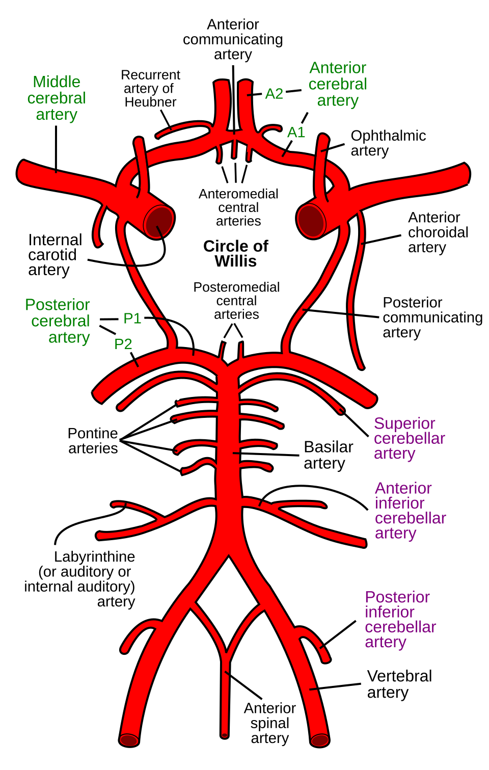
| Anterior choroidal artery | Major supply |
| Anterior temporal branches | Supplemental supply |
| Posterior cerebral artery branches | Supplemental supply |
Venous Drainage
| Vein |
|---|
| Deep middle cerebral vein |
| Basal vein |
| Temporal venous tributaries |
Development
The amygdala develops from both pallial and subpallial embryonic territories.
| Embryological Region | Adult Nuclei |
|---|---|
| Pallial amygdala | Lateral, Basal, Accessory Basal nuclei |
| Subpallial amygdala | Central and Medial nuclei |
This dual developmental origin contributes to the anatomical diversity of its nuclei.