Spinal Cord Anatomy
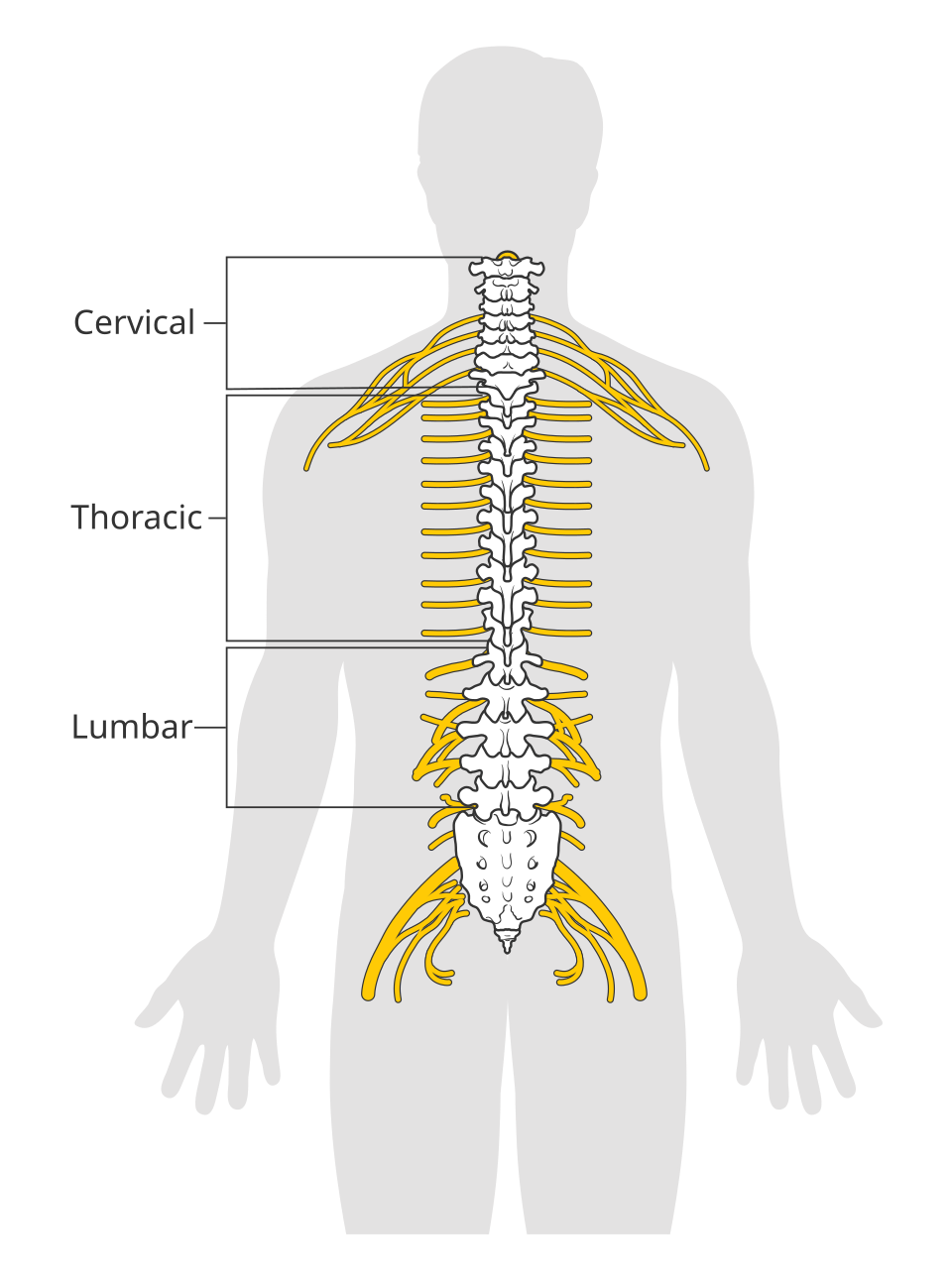
The spinal cord is the elongated portion of the central nervous system that connects the brain to the body. It transmits sensory information to the brain and motor commands to peripheral tissues.
- Superiorly continuous with the medulla oblongata at the foramen magnum.
- Terminates as the conus medullaris in adults.
- Continues inferiorly as the filum terminale.
- Inferior spinal nerve roots form the cauda equina.
Enlargements
Cervical Enlargement
- Spinal cord segments: C4-T1
- Supplies the upper limbs
Lumbosacral Enlargement
- Spinal cord segments: L1-S3
- Supplies the lower limbs
Meninges and Protective Spaces
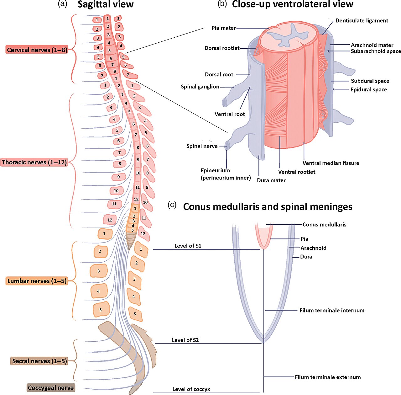
Three concentric meningeal layers encase the spinal cord:
- Dura Mater: A tough, fibroelastic outer tubular sheath extending from the foramen magnum to the S2 level, where it terminates as a blind sac.
- Epidural Space: Located between the dura and vertebral canal periosteum; contains adipose tissue and the internal vertebral venous plexus.
- Arachnoid Mater: A non-vascular intermediate membrane closely applied to the dura.
- Subarachnoid Space: Lies between the arachnoid and pia mater; contains cerebrospinal fluid (CSF) and major spinal vessels.
- Pia Mater: A vascular membrane directly adhering to the cord surface.
- Denticulate Ligaments: Lateral ribbon-like projections (21 pairs) of pia extending through the arachnoid to anchor the cord to the inner dural wall.
Cross-Sectional Architecture
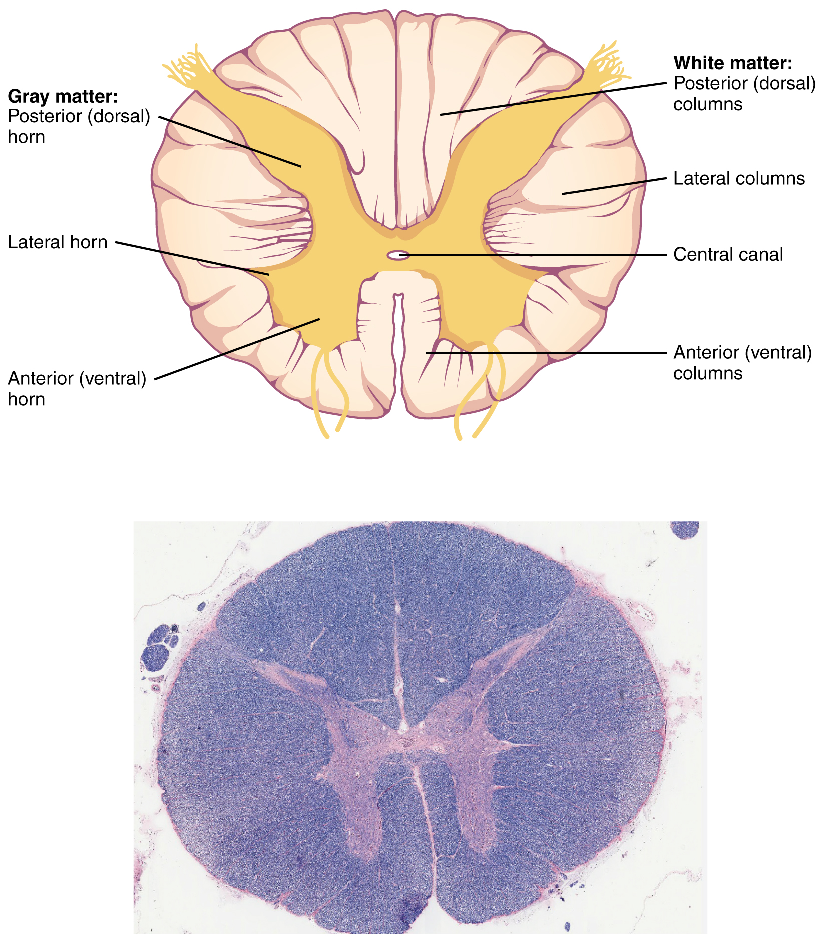
A transverse section reveals an inner H-shaped gray matter core surrounded by outer white matter funiculi, divided symmetrically by the anterior median fissure and posterior median sulcus.
Gray Matter (Neuronal Cell Bodies)
- Posterior (Dorsal) Horn: Receives somatic and visceral sensory inputs via dorsal root ganglion fibers. Contains Rexed laminae I through VI.
- Lateral Horn: Located exclusively in spinal segments T1-L2 (sympathetic preganglionic cell bodies) and S2-S4 (parasympathetic preganglionic cell bodies).
- Anterior (Ventral) Horn: Houses alpha and gamma lower motor neurons supplying somatic skeletal muscle fibers. Contains Rexed laminae VII through IX.
- Central Canal: Centrally located microchannel lined with ependymal cells, continuous superiorly with the fourth ventricle of the brainstem.
White Matter (Myelinated Axons and Tracts)
- Posterior Funiculus (Dorsal Columns):
- Fasciculus Gracilis: Medial tract carrying conscious proprioception, vibration, and discriminative touch from lower body segments (below T6).
- Fasciculus Cuneatus: Lateral tract carrying sensory fibers from upper body segments (T6 and above).
- Lateral Funiculus:
- Lateral Corticospinal Tract: Principal descending tract for voluntary motor execution.
- Spinothalamic Tract (Anterolateral System): Ascending nociceptive, thermal, and crude touch modalities.
- Dorsal and Ventral Spinocerebellar Tracts: Non-conscious proprioception routed to the cerebellum.
- Anterior Funiculus:
- Anterior Corticospinal Tract: Descending motor pathways for axial and proximal musculature.
- Vestibulospinal and Reticulospinal Tracts: Descending pathways regulating balance, posture, and autonomic muscle tone.
Segmental Organization and Vasculature
| Anatomical Parameter | Specific Characteristics |
|---|---|
| Spinal Nerve Count | 31 bilateral pairs total: 8 cervical (C1-C8), 12 thoracic (T1-T12), 5 lumbar (L1-L5), 5 sacral (S1-S5), 1 coccygeal (Co1). |
| Root Morphology | Dorsal root (sensory afferents, contains dorsal root ganglion) joins ventral root (motor efferents) to create a mixed spinal nerve. |
| Arterial Network | Single anterior spinal artery supplies anterior two-thirds of cord cross-section; paired posterior spinal arteries supply posterior one-third. |
| Radicular Reinforcement | Segmental medullary arteries reinforce longitudinal trunks; includes the Artery of Adamkiewicz (great anterior radicular artery). |
| Venous Drainage | Longitudinal anterior and posterior spinal veins drain via radicular veins into Batson internal vertebral venous plexus (valveless system). |
References
- "Diagram of the spinal cord" by Cancer Research UK, via Wikimedia Commons, licensed under CC BY-SA 4.0.
- Spinal cord details by Sheryl Tan, Faull L, and Curtis A, via Wikimedia Commons, licensed under CC BY 4.0.
- Spinal cord cross section. Illustration by OpenStax College via Wikimedia Commons (CC BY 4.0).