Photoreception
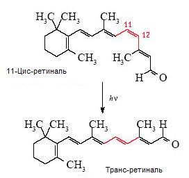
Photoreception begins in the membranous discs (lamellae) of photoreceptor outer segments, where light triggers the phototransduction cascade. In rod cells, the visual pigment rhodopsin consists of a 7-transmembrane G-protein-coupled receptor (opsin, composed of 348 amino acid residues) covalently linked to the chromophore 11-cis-retinal at lysine residue 296 (Lys296) via a protonated Schiff base. Cones use homologous opsins tuned to distinct spectral wavelengths. Upon absorbing a photon, 11-cis-retinal isomerizes into all-trans-retinal, inducing conformational transitions in opsin to form the catalytically active intermediate metarhodopsin II. (All-trans-retinal subsequently dissociates and is reduced to all-trans-retinol, or Vitamin A, to be recycled through the retinoid visual cycle).
Metarhodopsin II activates the heterotrimeric G-protein transducin (Gt), prompting its alpha-subunit to exchange GDP for GTP. Activated G(alpha t)-GTP then releases the inhibitory constraints on cGMP phosphodiesterase 6 (PDE6). Activated PDE6 rapidly hydrolyzes cyclic GMP (cGMP) to 5’-GMP at a rate exceeding 10^3 molecules per second.
Under dark conditions, elevated cGMP levels keep cyclic nucleotide-gated (CNG) cation channels open, driving a steady inward flux of Na+ and Ca2+ (the “dark current”) that maintains the photoreceptor in a depolarized state (~ -40 mV) with tonic glutamate release. The rapid depletion of cGMP by PDE6 closes these channels, leading to cell hyperpolarization and a subsequent reduction in glutamate release onto downstream bipolar and horizontal cells.
Recovery and adaptation occur through coordinated shut-off mechanisms:
- Pigment Inactivation: Rhodopsin kinase (GRK1) phosphorylates active metarhodopsin II, allowing arrestin-1 to bind and prevent further transducin activation.
- PDE Deactivation: Transducin’s intrinsic GTPase activity–substantially accelerated by the GTPase-activating protein complex RGS9-1/Gbeta5/R9AP–hydrolyzes GTP to GDP, releasing PDE6 back into its inactive state.
- Calcium-Mediated Adaptation: The closure of CNG channels halts Ca2+ entry while Na+/Ca2+,K+ exchangers continue extruding it. The resulting drop in cytosolic Ca2+ disinhibits guanylyl cyclase (via GCAPs), stimulating rapid cGMP resynthesis to reopen CNG channels and reset retinal sensitivity.